The Under the Coverslip competition pulls back the curtain on some of the beautiful and colour-splendid sights that can be found only by staring down the lens of the microscope.
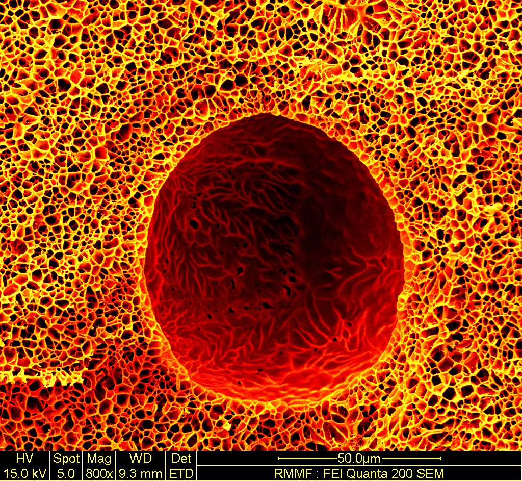
‘Microscopic Spaghetti Bubble’, by RMIT University PhD student Mitchell Boyd-Boss, was the first prize winner. Image depicts microscopic bubbles in the context of tissue engineering.
There’s a running joke among science students that no matter how many years of tertiary study we have, instructional videos we watch, or knobs we adjust ever so slightly – focusing a microscope and to reveal the meaning in the smudge on the slide is a dark and mystical unseen art.
It’s because of this that the submissions to the Under The Coverslip Competition, a photographic art competition for research scientists, are all the more impressive. What started two decades ago as a small group of students sharing photos from their research has grown into a nation-wide competition, attracting more than a hundred submissions from across Australia. Held by the Students of Neuroscience and Anatomy (SONA) society within the School of Biomedical Sciences at the University of Melbourne, it is the first and largest competition of its kind in Australia.
Jennifer Keller, SONA President, explained that “[the competition] is built on the notion that science is more than just graphs and figures; that there is undiscovered beauty beneath the microscope that can be appreciated by everyone.” And looking at some of the image submissions and photographs from competitions past, the beauty is clear to see.

‘Aboriginal Art’, by Monash University PhD student Nazia Tabassum came in second place. Image shows close-up mouse skin, showing sebaceous glands, hair follicle bulbs, distinct layers of skin, and other details.
The competition receives photos from a wide range of disciplines. “Microscopy is the backbone of so much research”, said Anna Wang, SONA Vice-President. As a result, the competition showcases many different subjects “ranging from the macroscopic surgical microscopy, to microscopic immunohistochemistry, to electron microscopy”.
The images exhibited would ordinarily be generated as a by-product of research. Under the Coverslip provides a platform for sharing these photographs usually exclusive to academia with the general public. The applicants are passionate researchers who are scientific experts on the subject of their art, who have access to the resources and opportunities that make the photos possible.

‘Living on the edge – supporting life with spiky micro-cones’ by University of South Australia PhD student Argha Chakraborty, was the third place winner. Image depicts an immune cell encountering a surface, reaching out to grip the micro-cone posts.
Many of the photographs fall under the category of accidental art, which Wang said is a major characteristic of the competition. “The winner this year took an image of their material used to grow neurons. Unexpectedly they came across a microscopic bubble that was not supposed to be in there, but it made a fantastic image.” The winning image, Microscopic Spaghetti Bubble, can be seen at the top of this article.
Another entrant had a similar story: “I knew the protein marker I was looking for was present in a completely unrelated part of the tissue, so out of curiosity I had a look. I found it was quite beautifully stained despite not needing an image of it for my research.”
Attendees can also hear the stories behind the photographs, as many of the entrants attend the exhibition themselves. Wang said that “this makes science more approachable [by] challenging the perception of science as being devoid of artistic flair.” Another aim of the competition is to “bring scientists from a broad range of fields together, allowing a greater appreciation of others’ research” within the scientific community”.

‘Brain based universe’ by University of Melbourne PhD student Fatemah Daemi took home the People’s Choice award. Image shows protective glial cells from the middle section between the hemispheres of a mouse brain. Astrocytes, named for their star-like shape, are labelled with a green marker and Microglia are marked with red.
In previous years, the exhibition went for one evening only. This year it kicked off on 9 November with the prizegiving, with the images on display from 12 to 15 November. The images have also appeared as part of Melbourne’s White Night, in Wang’s words “highlighting the public’s fascination with science and how beautiful it can be”. A collaboration with Science Gallery Melbourne in their upcoming permanent space is on the horizon for 2022.
Wang is optimistic that, by making science more accessible to the public, the Under the Coverslip competition will give back to research itself. “Hopefully by seeing scientists and science as interesting and approachable, we can encourage more support for research and more understanding of why it must be done. To reveal what happens behind the scenes may remove the trepidation some may feel towards science.”
Edited by Deborah Kane and John Back